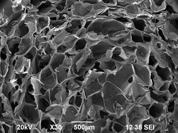
The team describes the alginate-based hydrogels as osteochindral tissue model
The design of an appropriate microenvironment for stem cell differentiation constitutes a multitask mission and a critical step toward the clinical application of tissue substitutes. With the aim of producing a bioactive material for orthopedic applications, a transforming growth factor-β (TGF- β1)/hydroxyapatite (HA) association within an alginate-based scaffold was investigated. The bioactive scaffold was carefully designed to offer specific biochemical cues for an efficient and selective cell differentiation toward the bony and chondral lineages. Highly porous alginate scaffolds were fabricated from a mixture of calcium cross-linked alginates by means of a freeze-drying technique. In the chondral layer, the TGF in citric acid was mixed with an alginate/alginate-sulfate solution. In the bony layer, HA granules were added as bioactive signal, to offer an osteoinductive surface to the cells. Optical and scanning electron microscopy analyses were performed to assess the macro-micro architecture of the biphasic scaffold. Different mechanical tests were conducted to evaluate the elastic modulus of the grafts. For the biological validation of the developed prototype, mesenchymal stem cells were loaded onto the samples; cellular adhesion, proliferation and in vivo biocompatibility were evaluated. The results successfully demonstrated the efficacy of the designed osteochondral graft, which combined interesting functional properties and biomechanical performances, thus becoming a promising candidate for osteochondral tissue-engineering applications.
L. Coluccino, P. Stagnaro, M. Vassalli, S. Scaglione (2016): “Bioactive TGF-β1/HA alginate-based scaffolds for osteochondral tissue repair: design, realization and multilevel characterization”. J Appl Biomater Funct Mater.
All Resources
Never stop learning!
Check publications from the team, protocols, and useful information to boost your research and get into organ on chip technology!


